


Coxarthrosis of the hip joint is a degenerative-dystrophic process that occurs in the articular joint of the femoral head and the pelvic acetabulum. The disease is more typical for middle-aged and elderly people, although it can also occur in young people, including children. Most often, its development is preceded by injuries, as well as a number of pathologies of an inflammatory and non-inflammatory nature, and pain and stiffness of movements become the main signs of a degenerative-dystrophic process in the hip joint. In its development, the disease goes through several stages, and if in the early stages it can be dealt with conservatively, then in the last stages, the treatment of coxarthrosis of the hip joints is effective only by surgery. Otherwise, the pathology will lead to severe disorders or even complete immobilization.
What is coxarthrosis of the hip joint and the mechanism of its development
Coxarthrosis, also called osteoarthritis and deforming arthrosis, is a complex disease of the hip joints (HJ), accompanied by progressive destruction of cartilage. Over time, this leads to deformation of the surfaces of adjacent bones, as well as the formation of bone growths on them, called osteophytes.
According to statistics, coxarthrosis accounts for about 12% of all diseases of the musculoskeletal system. In terms of frequency of occurrence, it is second only to gonarthrosis of the knee joint, but the risks of getting disability with it are much higher.
The two hip joints are the largest joints in the body. Each of them is formed by the femur bone and the acetabulum of the pelvis. The femoral head is located in the cup-shaped recess of the pelvic bone and moves freely in different directions. This structure of the joint makes it possible to flex and unbend, adduct and abduct, and also rotate the thigh.
To prevent movement from causing discomfort, the surfaces of the bones that touch each other are covered with an elastic layer called hyaline cartilage. It is he who allows the femoral head to easily slide in the acetabulum. Also, hyaline cartilage provides stabilization and cushioning of the hip joint during movements.
The entire joint is immersed in a kind of case called the articular capsule. It contains the synovial membrane that synthesizes synovial fluid. It is she who lubricates the surface of the cartilage, ensures the flow of water and nutrients into it, i. e. , is responsible for maintaining the normal structure of the cartilage tissue.
Above the joint capsule is a group of femoral and pelvic muscles, with the help of which the joint is set in motion. The hip joint is also surrounded by a group of ligaments that ensure the stability of its position within the physiological boundaries.
Since the hip joint is subjected to heavy loads every day, it is prone to rapid wear and injury. The risk of such changes significantly increases the effect of a number of unfavorable factors that are practically inevitable in the modern world, but they will be discussed below. This explains the high prevalence of coxarthrosis.
As a result of the influence of negative factors, there is a violation of the production of synovial fluid. Gradually, its quantity decreases, and its qualitative composition also changes: it becomes viscous, thick and is no longer able to fully nourish the cartilage. This leads to acute nutritional deficiencies and progressive dehydration of the hyaline cartilage. As a result of such changes, the strength and elasticity of cartilage tissue decreases, it exfoliates, cracks and decreases in volume. All this prevents the smooth sliding of the femoral head in the acetabulum of the pelvis, which leads to the appearance of signs of hip coxarthrosis.
Gradually, the interarticular gap narrows, increased friction occurs between the articulating bone surfaces, and the pressure of the bones on the hyaline cartilage increases. This leads to even greater injury and wear, which cannot but affect the biomechanics of the hip joint and the well-being of a person.
The failure of the hip joint negatively affects not only the biomechanics of the lower extremities, but also the entire locomotor apparatus. This often results in disability.
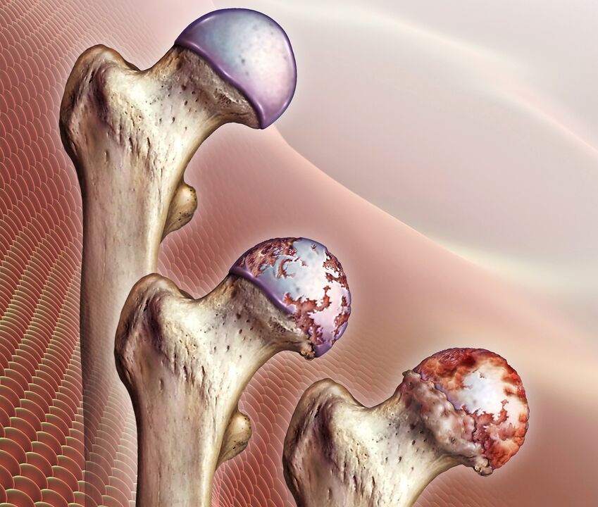
As pathological changes progress, the hyaline layer gradually disappears completely, which leads to the exposure of bone surfaces and a critical increase in the load on the bone joint. During movements, the femoral head is no longer covered by anything and rubs directly against the surface of the pelvic acetabulum. Besides the fact that it seriously limits mobility and causes unbearable pain, the bones press against each other, simultaneously flattening.
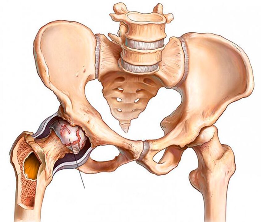
As the articulation bones deform, bone outgrowths (osteophytes) form on their surface. They can have sharp edges and seriously injure the surrounding muscles. This provokes the occurrence of severe pain in the groin, legs and buttocks. Therefore, the patient unconsciously tries to spare the affected hip joint and avoid movements in it. The lack of adequate load on the muscles leads to their gradual atrophy, which further exacerbates mobility problems. This results in lameness.
Reasons for development
Coxarthrosis of the hip joint can be primary or secondary. In the first case, the reasons for its development cannot be found, i. e. , the disease develops on its own for no apparent reason. Secondary coxarthrosis is the result of a number of changes in the state of the musculoskeletal system or lifestyle features, in particular:
- injuries of the hip joint, including bone fractures, dislocations, bruises, sprains or ruptures of the surrounding ligaments, chronic microdamages, etc. ;
- exhausting physical work;
- sedentary lifestyle;
- obesity;
- chronic infectious processes in the body;
- rheumatoid arthritis, gout, tendonitis, bursitis;
- endocrine diseases, metabolic and hormonal disorders, including diabetes mellitus;
- congenital malformations of the hip joint (dislocation, dysplasia);
- aseptic necrosis of the femoral head;
- pathologies of the spine of various kinds;
- genetic predisposition;
- addiction to smoking.
In the vast majority of cases, the development of hip joint coxarthrosis is due to inevitable age-related changes, and the presence of other factors from among the above only increases the risk of its occurrence and increases the rate of progression.
Symptoms and degrees
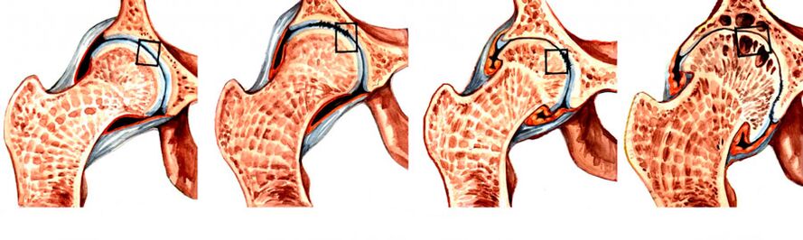
During coxarthrosis, 4 degrees of development are distinguished, of which 1 is the easiest. Initially, the disease may be asymptomatic or manifest as mild pain. More often they occur after intense physical exertion, a long walk or at the end of a busy day. At the first stages of the development of the disease, discomfort is usually attributed to fatigue and is regarded as the norm. Therefore, extremely rarely, coxarthrosis of hip joint is diagnosed at the 1st stage of development.
Perceptible signs of coxarthrosis begin to appear at the 2nd stage of its progression, when the joint space narrows by almost half, and the femoral head is displaced and deformed. With the transition to the 3rd stage, the pains become unbearable and can disturb a person even at night, they tend to radiate to the hips, shins, groin and buttocks. Since the joint space is already practically absent, and multiple osteophytes are formed on the bone surfaces, independent movement in such situations is impossible. Therefore, patients are forced to use a cane or crutches.
So, the main symptoms of coxarthrosis of the hip joint are:
- Mobility restrictions - initially, patients may notice the appearance of difficulties in performing rotational movements of the leg, but over time, morning stiffness and swelling of the HJ join them. Because of them, a person needs several minutes to warm up and, so to speak, walk around in order to restore a normal range of motion. Gradually, it becomes more and more difficult for the patient to perform leg movements.
- A characteristic crunch - occurs when walking, as well as flexion or extension of the hip joint. It is a consequence of the friction of bone surfaces against each other and with coxarthrosis is accompanied by sharp or dull pain.
- Pain syndrome - initially pains appear after physical exertion and subside somewhat after a long rest. An acute attack can be provoked by weight lifting or hypothermia, since coxarthrosis is often complicated by the addition of inflammation of the synovial membrane. As the disease progresses, the pain becomes more frequent, lasts longer, and gets worse.
- Spasm of the thigh muscles - is a consequence of pinching of the nerves and weakening of the ligamentous apparatus, so the muscles spasm compensatory to keep the head of the femur in the acetabulum. Also, muscle spasm can be provoked by the addition of synovitis.
- Lameness - occurs in the last stages of the development of the disease, since the deformation of the bone surfaces provokes the appearance of contracture of the flexor muscles. Therefore, a person cannot fully straighten the leg and keep it in this position. Also, the patient may involuntarily limp to transfer weight to the healthy half of the body, as this helps to reduce the intensity of pain.
- Shortening of the leg - observed with coxarthrosis of the 3rd degree. The leg on the side of the affected hip joint may be shortened by 1 cm or more as a result of narrowing of the joint space, decreased muscle tone, and flattening of the femoral head.
At the last stage of development, the femoral head fuses with the acetabulum, which leads to complete immobilization of the leg and disability.
At the same time, degenerative-dystrophic changes can be observed in one hip joint or both. Accordingly, characteristic symptoms will be observed either on one side or on both at once, but in the latter case, their severity on the left and right may differ.
Diagnostics
The doctor can suspect the presence of coxarthrosis of the hip joint based on the patient's complaints, external examination and the results of functional tests. Be sure to measure the length of the legs during a visual inspection. For this, the patient is asked to stand up and straighten his legs as much as possible. The measurement is taken between the anterior axis of the pelvic bones and any bony structure of the knee, ankle or heel. But if both hip joints are simultaneously affected by coxarthrosis, the data obtained will be uninformative.
But since symptoms typical of coxarthrosis can accompany a number of other inflammatory and non-inflammatory diseases, instrumental examination methods are mandatory for the patient to accurately diagnose the pathology. It could be:
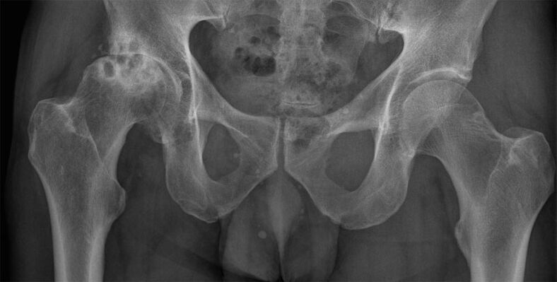
- CT or X-ray of the hip joint - the images show destructive changes in it, narrowing of the joint space, the formation of osteophytes and deformation of the bone surfaces;
- MRI is the most informative examination method that allows you to accurately assess the nature of changes in cartilage structures, ligaments, and the nature of blood circulation in the hip area.
Patients are also assigned laboratory tests to assess their general health and detect diseases that could cause coxarthrosis. It:
- UAC and OAM;
- blood chemistry;
- rheumatic tests;
- puncture of the hip joint with a biochemical study.
The task of diagnosis is to differentiate hip coxarthrosis with gonarthrosis (damage to the knee joint), as well as radicular syndrome that occurs with osteochondrosis, as well as protrusions and hernias of the intervertebral discs. Also, the symptoms of coxarthrosis may resemble manifestations of trochanteric bursitis and an atypical course of ankylosing spondylitis, which requires a full examination in order to find out the true causes of pain and mobility restrictions.
Conservative treatment
Conservative treatment of hip coxarthrosis is effective only at the initial stages of the disease. It is selected for each patient individually and may include a whole range of different methods, each of which will complement the others. Therefore, as part of the treatment of coxarthrosis of the hip joint, patients can be prescribed:
- drug therapy;
- exercise therapy;
- physiotherapy;
- plasmolifting.
In order for conservative treatment to be effective, patients need to eliminate the effect of a number of factors that contribute to the development of hip coxarthrosis. If you are overweight, it is very important to reduce it as much as possible. This will reduce the load on the affected joint and the risk of progression of the degenerative-dystrophic process.
You should also give up smoking and normalize the mode of physical activity, avoid overload, but do not sit all the time. To prevent further destruction of the hip joint, it is recommended to wear special bandages and orthoses. They provide a secure fixation of the joint and support it during movement.
Medical treatment
The nature of drug therapy is selected strictly individually. In most cases, patients are prescribed:
- NSAIDs - drugs that simultaneously have analgesic and anti-inflammatory effects (available in the form of tablets, injections and topical agents);
- corticosteroids - drugs with a powerful anti-inflammatory effect, which are prescribed if NSAIDs do not give a pronounced effect;
- chondroprotectors - contribute to the activation of cartilage tissue regeneration processes, but their effectiveness has not been proven;
- muscle relaxants - drugs that reduce muscle tone and eliminate spasms, which is necessary when spasming certain muscles or groups against the background of severe pain;
- preparations to improve blood circulation - are most often used in the form of injection solutions and help to improve the trophism of the tissues surrounding the joint;
- group B vitamins - are shown to normalize the transmission of nerve impulses, which is especially important when nerves are compressed by deformed bone structures.
For acute pain that cannot be eliminated with the help of tablets, intra-articular or periarticular blocks can be performed on patients. They are carried out exclusively by qualified health workers in a medical institution and involve the introduction into the joint cavity or directly the area around it of anesthetic solutions with corticosteroids.
exercise therapy
Therapeutic exercise is an effective method of dealing with a decrease in muscle tone and limitation of mobility. Thanks to a properly selected set of exercises, it is possible to increase the range of motion and reduce the severity of pain. They also prevent muscle atrophy and help eliminate spasms if coxarthrosis is accompanied by pinching of nerve fibers, which reflexively leads to spasm of individual muscles.
Exercise therapy classes can improve blood circulation in the area of the degenerative-dystrophic process. Due to this, the quality of the trophism of the diseased joint increases and the course of regenerative processes accelerates.
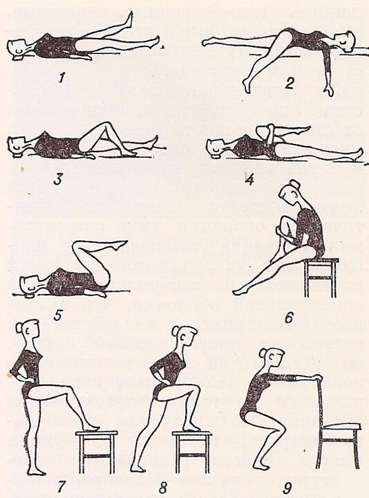
For each patient, a set of exercises should be developed individually by a specialist. At the same time, not only the degree of destruction of the hip joint is taken into account, but also the level of physical development of the patient.
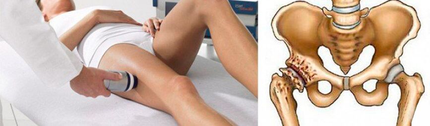
Physiotherapy
Physiotherapeutic procedures and massage have anti-inflammatory, analgesic, tonic, anti-edematous effect. Additionally, they help maintain normal leg muscle tone, preventing their atony and atrophy.
With coxarthrosis of the hip joint, courses of 10–15 procedures are prescribed:
- ultrasound therapy;
- magnetotherapy;
- laser therapy;
- electrophoresis;
- ultraphonophoresis;
- UHF;
- paraffin treatment.
Also, many patients are offered mud therapy. Such procedures have a positive effect only at the 1st stage of the development of coxarthrosis of the hip joint or during rehabilitation after surgical treatment. Thanks to therapeutic mud, it is possible to achieve an improvement in the quality of blood circulation and accelerate the restoration of the motor capabilities of the affected joint.
Plasmolifting
Plasmolifting or PRP-therapy is a procedure that involves the introduction of platelet-rich plasma of the patient's own blood into the cavity of the hip joint. This allows you to activate the processes of restoration of hyaline cartilage.
But, according to some scientists, such a procedure can cause the formation of malignant tumors. This point of view is based on the fact that plasmolifting promotes the formation of a large number of stem cells, the effect of which on the body has not yet been fully studied.
Surgical treatment of coxarthrosis of the hip joint
Despite the significant discomfort in hip joint, many seek medical help too late, when pathological changes in the joint reach 3 or even 4 degrees of severity, and the functionality is irreversibly depleted.
With advanced pathology, surgery is a necessary measure. Only a timely surgical intervention will help restore normal mobility and save the patient from excruciating pain, that is, to achieve a significant improvement in the quality of human life. No medicines, physiotherapeutic procedures, can restore severely destroyed cartilage. At best, painful intra-articular injections and drugs can reduce pain. But this will be a temporary phenomenon, after which the pain will return again with the same or even greater strength.
Indications for hip surgery are:
- disappearance of the interarticular space;
- persistent pain in the hip joint, not amenable to relief;
- critical mobility disorders;
- hip fracture.
Depending on the severity of joint destruction and bone deformity, patients can be offered various types of surgical treatment, namely:
- arthrodesis;
- endoprosthesis;
- osteotomy.
Arthrodesis
Arthrodesis is an affordable operation that involves a strong fixation of the articular bones with metal plates. The result is complete immobilization of the joint. Therefore, with the help of arthrodesis, it is possible to correct only the supporting function of the leg, eliminate pain, but it is not necessary to talk about restoring mobility or a significant improvement in the quality of life.
Today, arthrodesis is practically not used, as it deprives a person of the opportunity to fully move around.
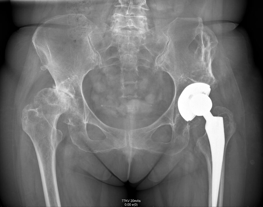
Endoprosthetics
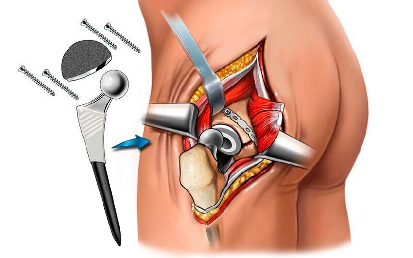
Endoprosthetics with arthroplasty is the only way to radically solve the problem of coxarthrosis of the hip joint with the restoration of all its functions and motor capabilities. This is a high-tech method for solving the problem of coxarthrosis, which allows you to completely forget about it for 15–30 years, as well as about pain and mobility restrictions. Thanks to the use of modern endoprostheses, it is possible to achieve full restoration of motor-support functions and provide the patient with a normal life.
The operation involves resection of the femoral head and part of its neck. Surgical preparation of the acetabular bed is also carried out, which involves the removal of osteophytes, the alignment of its surface and the resection of tissues that have undergone necrosis. Endoprosthetics can even be used to treat elderly patients with hip coxarthrosis.
The operation is performed under general anesthesia and takes about an hour. Depending on the severity of the degenerative-dystrophic process, the operation can be performed using one of the following methods:
- superficial - involves grinding the acetabulum and femoral head with further coating with smooth implants that replace the destroyed hyaline cartilage (the method is rarely used due to the possibility of inflammation in the periarticular tissues);
- unipolar - removal of the femoral head and its replacement with an endoprosthesis (used when cartilage is preserved on the surface of the acetabulum and only the femoral head is destroyed);
- bipolar - similar to the previous technique, differing only in the design of the endoprosthesis used, which has a lower coefficient of friction and provides smoother movements in the joint bed;
- total is the most effective and safest method for solving the problem of hip joint coxarthrosis, which involves a complete resection of the femoral head with the capture of part of its neck, as well as the acetabular fossa and replacing them with a full-fledged artificial articular joint.
Thus, patients may be recommended to install various types of endoprostheses. Most hip replacements are manufactured in the US and UK. For their manufacture, chemically and biologically inert metals are used: cobalt, chromium, titanium alloys. Often ceramics are also used. In most modern models, polymer pads are additionally used, which make it possible to provide natural shock-absorbing, stabilizing and sliding properties to artificial TBS.
When performing endoprosthetics, the success of the operation is almost 100%.
After the operation, antibiotics are prescribed to prevent the development of infectious complications, and the stitches are removed after 10 days. The size of the postoperative scar is approximately 8 cm. At the same time, the patient is discharged from the clinic. Rehabilitation after endoprosthetics is simple, but still requires physiotherapy, massage and exercise therapy.
osteotomy
Osteotomy is a surgical intervention that is a temporary measure before a cardinal replacement of the hip joint with an artificial endoprosthesis. The essence of the operation is to align the axis of the femur due to its intentional fracture. The resulting fragments are set in the most appropriate position, thereby slightly unloading the diseased joint. As a result, it is possible to temporarily reduce the severity of pain and improve mobility.
Thus, hip coxarthrosis is a rather formidable disease that can completely deprive a person of the opportunity to move independently. It progresses for a long time, and its symptoms, especially in the early stages, are often perceived by patients as a normal condition after physical exertion. But it is precisely in this that the insidiousness of the disease lies, because only at the initial stage of its development it can be dealt with in a non-surgical way. But if the degenerative-dystrophic process has already completely destroyed the hyaline cartilage and led to the exposure of the surfaces of the bones, and even more so to their flattening, only surgery can help the patient. Fortunately, the modern level of medicine and surgery, in particular, makes it possible to achieve a complete restoration of the normal state of the hip joint and its functions.