


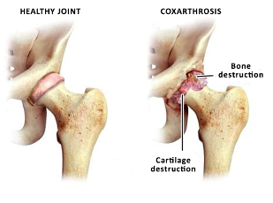
Coxarthrosis - degenerative disease that leads to destruction of the hip joint and has a chronic course. More common in older age groups. More common in women than men.
Onset is gradual and progresses slowly. May affect one joint or both. Is the most common type of osteoarthritis.
Why is the illness?
Osteoarthritis in some patients is accompanied by the natural aging process is the degeneration of the tissues of the hip joint. Its appearance is influenced by such factors:
- reduced tissue nutrition;
- congenital anomaly of the hip joint, in particular, dysplasia;
- trauma pelvic;
- post-infectious hip;
- aseptic necrosis of the head of the hip joint;
- Perthes disease (osteochondropathy).
Unfortunately, to determine the cause of the disease is not always possible and pathology of the hip joint is called idiopathic coxarthrosis - that is so, the cause of which is not established. This is the incentive for continuous study of the problem. Scientific works in this field and the doctors came to the conclusion that a higher risk of osteoporosis observed in the following patients:
- Hereditary predisposition to pathology. Patients whose parents suffered from diseases of cartilage and bone, in most cases, you will also have a such problems;
- Overweight. Significant body weight is the load on the joints that are regularly subjected to mechanical work;
- Metabolic disorders, diabetes. This leads to poor supply of oxygen and nutrients in joint tissue, causing them to lose their properties.
Knowing the main risk factors of the disease, and plan preventive measures to prevent it.
How to recognize the pathology of the hip joint?
Symptoms of osteoarthritis depends on the anatomical features of the musculoskeletal system, causes of the pathology and stage of the process. Consider the main clinical manifestations:
- painful joints;
- irradiation of pain in the knee, thigh, groin;
- stiffness of movement;
- limited mobility;
- violations walk, limp;
- the decrease in the mass of thigh muscles;
- shortening of the affected limb.
The clinical picture corresponds to the internal changes in the tissues of the joint. The symptoms increase gradually and in the early stages, the patient is not paying them enough attention. This is dangerous, because in the beginning of the treatment process brings a greater effect.
Clinical and radiological degree of osteoarthritis
Listed below are symptoms specific to each degree.
- 1 degree. The patient experiences intermittent pain and discomfort. Discomfort bother you after exercise, a long position in a static pose. Pain localized in the area of the joint and passes after rest. At this stage of the process is not impaired gait and no shortening of the leg. The changes seen on radiographs - joint space narrowing, there are osteophytes (bony growths).
- 2 the degree. Increases the intensity of the pain, it can occur during rest and radiates to nearby areas of the body. Appears limp after the man had walked or surge. Limited range of motion in the joint. In parallel, a change of the x-ray pictures: displaced head of the femur, osteophytes grow on the inner and outer edges of the acetabulum.
- Stage 3. Pain becomes permanent, appears in the daytime and at night. Much worse gait appears a permanent limp. Drastically reduced motor function, atrophy of the leg muscles. changing the muscle tissue causes leg slightly pulled up and becomes shorter. This leads to a deformation of posture and curvature of the body. The radiograph at this stage of the process: the total narrowing of the gap between the surfaces of the joint, deformity of the femoral head, significant growth of osteophytes.
Diagnostic program when the disease
The main method of diagnosis - x-ray. It can be used to determine the presence of the disease and its stage. On radiography analyze the structure of joint on the subject of narrowing of the joint space, osteophytes, fracture of the head of the hip bone.
If there is a need to study the condition of soft tissue MRI is performed. It allows to study in detail the condition of the cartilage areas of the joint and the muscles of the hip region.
Modern methods and ways of treatment of coxarthrosis of the hip joint
Treatment of osteoarthritis can be conservative and surgical. Treatment of osteoarthritis is aimed at the following objectives:
- reducing the pain symptoms;
- restoration of motor activity;
- rehabilitation and rehabilitation;
- prevention of complications;
- improving the quality of life of the patient.
Initiation of treatment is modification of risk factors. To do this, the doctor recommends the following actions:
- normalization of body weight;
- avoiding harmful habits;
- nutrition;
- normalization of physical activity;
- balanced drinking regime;
- a healthy sleep.
Conservative treatment are: drug and non-drug. Drug treatment includes nonsteroidal anti-inflammatory drugs, analgesics, chondroprotectors. They reduce inflammation in the tissue, eliminates swelling and soreness, restore range of motion and improve the condition of cartilage.
Non-drug treatment includes, among other things, massage the affected area. It stimulates the muscles that oppose their degeneration and prevention of shortening of the limb. Full and professional massage stimulates blood flow in the area of the joint, and this, in turn, leads to normalization of metabolism in tissues. Please note that massage is not always helpful when coxarthrosis - it is conducted only between exacerbations and at certain stages of the process. Assign it to your physician may, recommend the massage techniques, the multiplicity of treatments and the duration of the course.
Obligatory condition of treatment is physiotherapy. Is prevention of contractures and progression of the disease. Exercises should be performed daily, only then will they have an effect. Exercises are chosen on an individual basis and is prescribed by a doctor. Exercises improve overall health, reduce the risk of emotional disorders, strengthen the powers of the body.
Physical therapy is another method that applies in coxarthrosis. It may be mud, medical baths and showers, magnetotherapy. Used electro - and phonophoresis of medicinal substances.
If these methods of treatment brought no effect or were applied late - surgical treatment is required.
Surgical intervention with coxarthrosis
Surgical treatment is used with the ineffectiveness of conservative methods. This is especially true for late diagnosis. Modern operational techniques and high-quality equipment operating allow to restore the structure and function of the joint, to restore human range of motion and a normal quality of life. The most effective method of surgical treatment is arthroplasty.
The indications for surgery are:
- coxarthrosis 2-3 degrees;
- the lack of effect of therapy;
- total restriction of movement, walking.
Contraindications, which do not allow to perform the operation:
- decompensated renal, heart, liver;
- mental illness;
- the acute phase of the inflammatory process in the body.
For this purpose, the preoperative diagnosis. However, if it is possible to adjust the condition of the patient preparing for surgery and after that the intervention.
The operation involves removing the affected tissue and the prosthesis. There are different models of implants. Different methods of fastening in the bone – cement and cement-free, the material from which the endoprosthesis. Of all the features of the prosthesis and the intricacies of surgical intervention can I get information on consultation with the attending physician.
The recovery period after surgical treatment
From the first day after surgery, rehabilitation is carried out under the supervision of a physician. First, she is to perform passive movements, then the load is increased gradually. Walking in the first time is allowed only with crutches, allowed the seat and squat.
Of course, the first time after the operation, there are restrictions on the loads. Do not be afraid - because without operation these limits would be preserved to the end of life. A decrease in physical activity after surgical treatment, it is necessary to strengthen the position of the prosthesis, restoration of the integrity of bones, healing wounds. Within 2 months should be excluded the sports activities physical exercise on the joint, a long walk and some exercise. After complete recovery, the person returns to normal life, can do sports and outdoor activities.
The service life of the prosthesis: the majority of firms indicates a survival rate of about 90% for observation periods up to 15 years.